Thursday, September 25, 2014

Phytochemicals

What are phytochemicals?

Phytochemicals are non-nutritive plant chemicals that have protective or disease preventive properties. They are nonessential nutrients, meaning that they are not required by the human body for sustaining life. It is well-known that plant produce these chemicals to protect themselves but recent research demonstrate that they can also protect humans against diseases. There are more than thousand known phytochemicals. Some of the well-known phytochemicals are lycopene in tomatoes, isoflavones in soy and flavanoids in fruits.

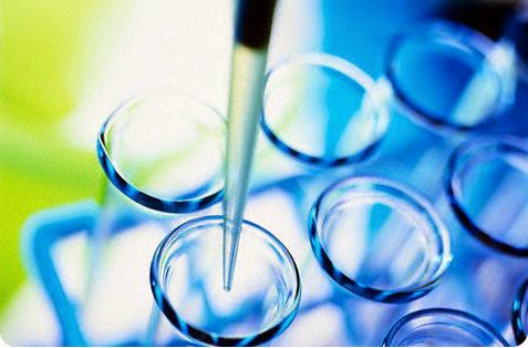
What is Phytochemical Screening?

It is a process of tracing plant constituents. For example you want to found out if a certain plant contains alkaloids (a plant constituent) then, you will be performing a phytochemical screening procedures for alkaloids (in this case mayer's and Wagner's test). There are general plant constituents that can be performed with a standard test.


What are the different reagents used in Phytochemical Screening? How are these prepared?


REAGENT-  is a "substance or compound that is added to a system in order to bring about a chemical reaction, or added to see if a reaction occurs." Although the terms reactant and reagent are often used interchangeably, a reactant is more specifically a "substance that is consumed in the course of a chemical reaction". Solvents, although they are involved in the reaction, are usually not referred to as reactants. Similarly, catalysts are not consumed by the reaction, so are not described as reactants.

-Mayer’s Reagent
(Alkaloid)

Procedure:
0.4 g of Mercuric Chloride is dissolved in 15 ml water and poured into a solution of 1.25 Potassium Iodide in 2.5 ml of water. Sufficient water was added to make 25 ml.

-Molisch’s Reagent
(Carbohydrates)
Procedure:
1.5 g of Naphthol was dissolved in 10 ml of ethanol

-Acetic acid
Ferric Chloride
(Glycosides)

Procedure:
Few drops of ferric chloride was mixed with 2.5 ml of acetic acid

-Distilled Water
(Saponins)

Procedure:
Pure extract of substance was mixed with distilled water

-Distilled Water
Ferric Chloride
(Phenols)
Procedure:
Mix Distilled Water and Ferric Chloride

-Ferric Chloride solution
(Tannins)

Procedure:
Dissolve 13.5 g of Ferric Chloride in 10 ml water with 0.25 ml of concentrated Hydrochloric Acid Dilute to 100 ml


-Sodium Hydroxide Solution
(Anthocyanin)

Procedure:
Dissolve 0.4 g of Sodium Hydroxide in 10 ml of water.

-Nitric Acid
(Protein)

Procedure:
Mix Nitric Acid to the solution

-Sodium Hydroxide Solution
(Flavonoids)

Procedure:
Dissolve 0.4 g of Sodium Hydroxide in 10 ml water.


What are the indicators of the presence of each phytochemical?

Alkaloids
In Determinig the presence or absence of alkaloids, Mayer’s reagent test was used. It is a mixture of mercuric chloride solution and potassium iodide solution. Alkaloid is present if there is a formation of green or white precipitations.

Carbohydrates
To determine if carbohydrates were present, Molisch’s reagent is used. It was prepared by mixing naphthol and ethanol. Carbohydrates were present if there is a formation of red or purple when the extract was mixed with Molisch’s reagent.

Glycosides
In testing the presence of glycosides, acetic acid and ferric chloride was used. There is a presence of glycosides if the color of the mixture extract and reagent became blue-green.

Saponins
To test of Saponins were present on the extract, ditilled water was added and then chook for fifteen minutes. When there is a formation of foam or frothy bubbles on the mixture, it indicates that saponins were present.

Phenols
The picture shows that traces of Phenols were present in the extract. The brown extract changes into the green color.The formation of blue or green color indicates the presence of phenols.

Tannins
To determine the presence of tannins, ferric chloride solution was used. When there is a formation of dark blue or greenish black color indicates the presence of tannins.

Anthocyanin

The presence or absence of anthocyanin as determined by the use of sodium hydroxide solution., when there is a formation of blue or green precipitates. It indicates that anthocyanin Is present.

Protein
The formation of yellow, yellow-green or light green color when the extract was mixed with nitric acid indicates the presence of protein.

Flavonoids
Sodium Hydroxide solution was used to determine the presence of flavonoids. Formation orange of intense yellow color indicates its presence.
---------------------------------------------------
WHAT I LEARNED...
I learned what are Phytochemicals and what are the Reagents used in Phytochemical Screening and the indicators to know the presence of a certain Phytochemical.

WHAT I DID...
I Search for more details and information about Phytochemicals to gain more knowledge and to discover new things that can guide us in our Investigatory Project.

I CAN APPLY MY LEARNING TO...
I can use the information I got by using it as a guide in doing our Investigatory Project,

Thursday, August 21, 2014

Bacteria

What is a Bacteria?

Bacteria
Bacteria constitute a large domain of prokaryotic microorganisms. Typically a fewmicrometres in length, bacteria have a number of shapes, ranging from spheres to rods and spirals. Bacteria were among the first life forms to appear on Earth, and are present in most of its habitats. Bacteria inhabit soil, water, acidic hot springs,radioactive waste,and the deep portions of Earth's crust. Bacteria also live in symbiotic and parasitic relationships with plants and animals. They are also known to have flourished in manned spacecraft.

What are the different kinds of Bacteria and their Gram-Stain?

Gram-Positive

Cocci
Gram-positive anaerobic cocci (GPAC) are a heterogeneous group of organisms defined by their morphological appearance and their inability to grow in the presence of oxygen; most clinical isolates are identified to species in the genus Peptostreptococcus. GPAC are part of the normal flora of all mucocutaneous surfaces and are often isolated from infections such as deep organ abscesses, obstetric and gynecological sepsis, and intraoral infections. 

Bacillus

Bacillus is a genus of Gram-positive, rod-shaped (bacillus), bacteria and a member of the phylum Firmicutes. Bacillus species can be obliga
te aerobes (oxygen reliant), orfacultative anaerobes (having the ability to be aerobic or anaerobic). They will test positive for the enzyme catalase when there has been oxygen used or present.Ubiquitous in nature, Bacillus includes both free-living (non-parasitic) and parasitic pathogenic species


Gram-Negative

Spirillum

Spirillum in microbiology refers to a bacterium with a cell body that twists like a spiral. It is the third distinct bacterial cell shape type besides coccus and bacillus cells.Spirillum is the bacteria of a genus of Gram-negative bacteria (family Spirillaceae). There are two species, Spirillum volutans and Spirillum winogradskyi. The taxonomic position of Spirillum minus and Spirillum pulli is uncertain. Spirillum minus is associated with rat-bite fever, and Borrelia burgdorferi with Lyme disease.


Vibrios
Vibrio is a genus of Gram-negative bacteria possessing a curved rod shape (comma shape), several species of which can causefoodborne infection, usually associated with eating undercooked seafood. Typically found in saltwater, Vibrio spp. are facultative anaerobes that test positive for oxidase and do not form spores. All members of the genus are motile and have polar flagella with sheaths. Recent phylogenies have been constructed based on a suite of genes (multilocus sequence analysis).

Spirochaete
Spirochaetes (also spelled spirochetes) belong to a phylum of distinctive diderm (double-membrane) bacteria, most of which have long, helically coiled (corkscrew-shaped) cells. Spirochaetes are distinguished from other bacterial phyla by the location of their flagella, sometimes called axial filaments, which run lengthwise between the bacterial inner membrane and outer membrane in periplasmic space. These cause a twisting motion which allows the spirochaete to move about. When reproducing, a spirochaete will undergo asexual transverse binary fission. Most spirochaetes are free-living and anaerobic, but there are numerous exceptions.


What diseases are caused by these bacterias?


COCCI- It causes strep throat, some skin diseases and pneumonia, among many others. They can also cause gonorrhea, meningitis, and skin lesions.

BACILLUS- Escherichia coli is a rod-shaped bacteria that normally lives in your intestinal tract without causing disease. However, a few strains of E. coli do cause disease that's spread typically by eating or drinking contaminated food or water; a typical symptom is diarrhea. Corynebacterium diphtheriae, another rod-shaped bacterium, infects the respiratory tract and causes diphtheria. Diphtheria causes a thick coating on the back of the nose and throat, making it difficult to swallow or breathe, followed by swelling of the neck and potentially death. Bacillus anthracis is the bacteria that cause anthrax. This rod-shaped bacterium grows in long chains and can infect you through broken skin, ingestion or inhalation.

SPIRILLUM- Rat-Bite fever is one disease caused by spirilla minus (the minus doesn't really mean that much) my source is the CDC spirilla is split into sub groups like: treponema pallida, that causes syphilis Borrelia burgdorferi, that causes lyme disease my source is Milady's Standard Cosmetology

VIBRIOS- They typically live in aquatic environments. Vibrio cholera moves in a darting motion by a single flagellum, a whiplike structure, and is the bacteria that causes cholera. Cholera is an intestinal infection that causes severe diarrhea and dehydration, typically transmitted by drinking contaminated water. It is a very serious disease that can lead to death if not treated promptly.

SPIROCHAETE- Two well-known spirochetes that cause disease in humans are Treponema pallidum and Borrelia burgdorferi. Treponema pallidum causes the sexually transmitted disease syphilis. Infection typically begins as a single sore at the site of infection. Additional lesions or rashes can develop elsewhere on the body if left untreated. Borrelia burgdorferi is transmitted through the bite of an infected tick and causes Lyme disease. Infection with Borrelia burgdorferi causes a typical "bull's-eye" rash. If left untreated, it can affect your heart and nervous system and cause arthritis.

What are the differences between Gram-Positive and Gram-Negative Bacteria?

Gram-Positive

Gram-positive bacteria are classified as bacteria that retain a crystal violet dye during the Gram stain process. Gram-positive bacteria will appear blue or violet under a microscope, whereas Gram-negative bacteria will appear red or pink. The difference in classification is largely based on a difference in the bacteria's cell wall structure.
The following characteristics are generally present in a Gram-positive bacteria:
1. A very thick cell wall (peptidoglycan)
2. If a flagellum is present, it contains two rings for support (an additional two rings are not necessary, unlike in Gram-negative bacteria, because the thick cell wall is supportive enough)
3. Teichoic acids are present, which serve to act as chelating agents, and also for certain types of adherence

Gram-Negative

Gram-negative bacteria are those that do not retain crystal violet dye in the Gram staining protocol. Gram-positive bacteria will retain the dark blue dye after an alcohol wash, whereas Gram-negative bacteria do not. In a Gram stain test, a counterstain is added after the crystal violet, which colors all Gram-negative bacteria a red or pink color. The test itself is useful in classifying two distinctly different types of bacteria based on structural differences in their cell walls.

Many species of Gram-negative bacteria are pathogenic, meaning they can cause disease in a host organism. This pathogenic capability is usually associated with certain components of Gram-negative cell walls, in particular the lipopolysaccharide (also known as LPS or endotoxin) layer.

The following characteristics are displayed by Gram-negative bacteria:
1. Cell walls only contain a few layers of peptidoglycan (which is present in much higher levels in Gram-positive bacteria)
2. Cells are surrounded by an outer membrane of lipopolysaccharide outside the peptidoglycan layer
3. Porins exist in the outer membrane, which act like pores for particular molecules
4. There is a space between the layers of peptidoglycan and the secondary cell membrane called the periplasmic space
5. The S-layer is directly attached to the outer membrane, rather than the peptidoglycan
6. If present, flagella have four supporting rings instead of two
7. No teichoic acids are present
8. Lipoproteins are attached to the polysaccharide backbone whereas in Gram-positive bacteria no lipoproteins are present
---------------------------------------------------
WHAT I LEARNED...
I learned that Bacteria have different types like Cocci, Bacillus, Spirillum, Vibrio and Spirochete. I also found out the different illness caused by these bacteria. Lastly, I learned the difference between Gram-Positive bacteria and Gram-Negative Bacteria.

WHAT I DID...
I Search for more details and information about these bacteria to gain more knowledge and to discover new things that I can apply to my daily living.

I CAN APPLY MY LEARNING TO...
I can use the information I got by using it as a guide in doing our Investigatory Project and to our daily lives.